During scientific discovery, we encounter moments of fascination, not necessary all about science but also art.
Here, we intend to share some of our results that made us “wow”, as we call them “Science as Art“.
科学艺术,通常指在科学研究过程中,经意或不经意间得到的富有艺术感的图片结果。
让我们去找寻那些在科学实验中给予我们的“wow”时刻。。
Close-up of Zebrafish Eye
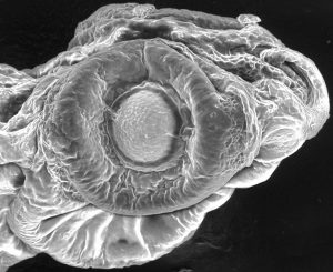 Zabrafish larvae (6 dpf) under scanning electron microscope. (by Sijie Lin)
Gentlemen in red bow tie
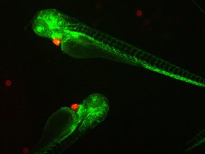 Double transgenic zebrafish larvae were created by crossing transgenic zebrafish strains flk-eGFP and clmc-dsRed. At Day 3 after birth, under a fluorescent microscope, the double transgenic larvae displayed the whole vascularture in green while heart in red. One fantastic creature for environmental safety assessment of chemicals, including engineered nanomaterials. (by Sijie Lin)
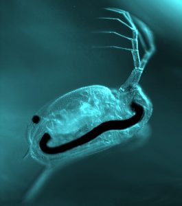
A hungry water flea
An aquatic organism called Daphnia magna with a gut full of black aggregates, as a result of exposure to multiwall carbon nanotube dispersion. (by Sijie Lin)
Hamlet (2009, 1st place, Science as Art competition, Clemson University)
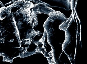 “Though this be madness, yet there is method in it”, (ACT II, Hamlet). This image was obtained as part of an experiment to study the interaction of nanoparticles with cellulose, the major component of bacteria and plant cell walls. The characters in the image above occurred unexpectedly in a sample of cellulose film, spin coated onto a microscope slide with a single wafer spin processor (Laurell Technologies). The amusing features, resembling the well-known characters in Hamlet, showed up when microcrystalline cellulose powder was dissolved in a solvent (9% LiCl:DMAC). They were likely residues of the cellulose and formed sporadically. Both science and art involve the ambitious quest of those moments when what one perceives suddenly becomes more than what was anticipated. This piece, in its own way, is a symbolic manifestation of beauty in the trash after a long day at the lab, during one of our numerous attempts in understanding the physics behind the biological study of life. The sample was platinum-coated and viewed at 15kV on a Hitachi S-4800 scanning electron microscope. (by Sijie Lin, Priyanka Bhattacharya, Tatsiana Ratnikova and Pu-Chun Ke)
Jewel (2009, Science as Art competition, Clemson University)
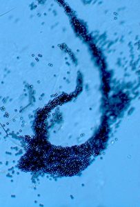 This image was obtained as part of an experiment to study the stability of microplastic beads in aquatic media, part of a research project on assessing the environmental impact of disposed plastics. The experimenters noticed in the experiment that micropastic beads readily self assembled in the media. The shape of this particular assembly reminded the experimenters of a beautiful cascade necklace gently drifting down the ocean water, while the glistening beads may be perceived as precious African stones. The image was obtained with a Zeiss A1 bright field microscope. (by Sijie Lin, Priyanka Bhattacharya, Ran Chen and Pu-Chun Ke)
Streaming, (2008, Science as Art competition, Clemson University)
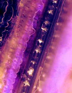 This image, resembling a guitar finger board, displays the remarkable uptake of multiwalled carbon nanotubes (bright agglomerates) by the vascular system (tubular structure) of a rice plant. It offers first-hand evidence of nanomaterials discharged into the food chain and illustrates the potential impact of nanotechnology on human health.
Understanding the impact of nanotechnology on the environment is of increasing urgency and great scientific importance. Our team combines advanced methodologies and techniques in both plant biology and biophysics to explore the ecological consequences of discharged nanomaterials. Rice plants, a major food crop species, have been found in this research to readily integrate multiwalled carbon nanotubes through their vascular systems (tubular structure in the image), the plant compartments for water and nutrient uptake. During transport in the vascular systems, nanotubes transform and aggregate into microsized particles (bright agglomerates). They further display an intriguing periodic pattern which largely coincides with the partitions of the plant stem. This image offers the first evidence of nanomaterial uptake by the food chain and their subsequent transport and transformation driven by diffusion and water convection. This image opens the door for our understanding of the fate of nanomaterials in ecological systems, and ultimately, the impact of nanomaterials on human health. (by Sijie Lin, Michelle L. Reid, Hong Luo & Pu-Chun Ke)
Chianti (2007, Science as Art competition, Clemson University)
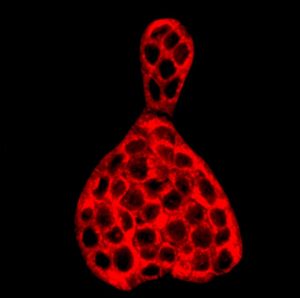 The fluorescence image was taken by a Zeiss LSM 510 confocal scanning microscope. It shows the cross sections of approximately fifty HT-29 colon cancer cells labeled with lipophilic dye Dil. Only the cell membranes fluoresce in vivid red while the exteriors and interiors of the cells display black. The envelopes of these cells resemble a Chianti bottle, the famed Italian red wine. Scale: 200X200 µm. (by Sijie Lin, Josh Mount, Xi Wang and Pu-Chun Ke)

